Brain - Center for Image Sciences
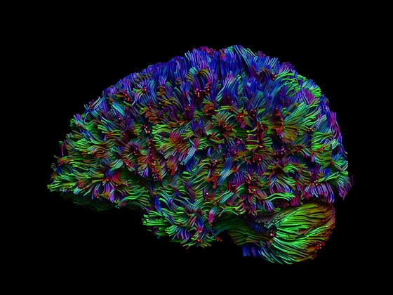
Detailed brain imaging has revolutionized our understanding of brain morphology and function in healthy subjects, aging populations and disease. This is true for CT, nuclear medicine and foremost MRI. MRI brain imaging uses the versatility of MRI with excellent soft tissue contrast to allow the assessment of brain morphology and function (fMRI) including for instance details of white matter tracts with diffusion tensor imaging (DTI).

Research participants

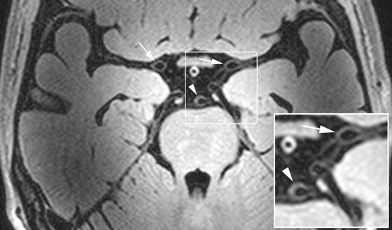
Arteries of the cerebral vessels in MRI
Patient care uitklapper, klik om te openen
Quantitative MRI methods for (regional) brain volume analysis and white matter lesion load are developed in populations studies and translated to daily clinical patient care to impact decision making in individual patients. The UMC Utrecht Center for Image Sciences has pioneered in the development and application over the full range of novel brain MR imaging methods noteworthy in the last decade in the field of ultra-high field MRI (7Tesla) and the use of innovative MRI brain methods in daily patient care.
Research field uitklapper, klik om te openen
The UMC Utrecht Center for Image Sciences is world leading in the development of innovative MRI brain imaging methods at high and ultra-high MRI field strength (3Tesla and 7Tesla MRI; 9.4 Tesla preclinical MRI). Newclinical and preclinical high-field imaging methods allow the detailed assessment of the brain tissue (micro infarcts) and the arterial vasculature including highly detailed vessel wall imaging. Other unique methods at ≥ 7Tesla are highly detailed MR imaging of the hippocampal subregions and MR spectroscopy (glutamate) and functional MRI of cortical gray matter layers and detailed assessment of the cerebrovascular reserve both with BOLD MRI and ASL MRI methods. Furthermore, together with the group of Prof. Max Viergever special focus is on the quantitative assessment of the brain with DTI analysis and the volumetric analysis of brain (sub) regions including the hippocampus, white matter lesions, microbleeds and smaller and larger infarcts.
Brain CT
For brain CT the UMC Utrecht has in depth expertise with CT perfusion and CT angiography methods which are systematically analyzed in a prospective cohort study of 1400 stroke patients (Dr. Birgitta Velthuis; Dutch Stroke Trial). Collaborations with clinical partners include the department of Neurology for several research fields: neurodegenerative diseases (Prof. Geert Jan Biessels), aneurysms (Prof. Gabriel Rinkel), cerebrovascular diseases (Prof. L Jaap Kappelle, Dr. Bart van der Worp) and ALS (Prof. Leonard van den Berg).
Other fields
Furthermore, collaborations in several other fields exists including: brain tumors research with the Neurosurgery department (Prof. Pierre Robe), carotid artery stenosis research with the department of Vascular Surgery (Dr. Gert Jan de Borst), epilepsy research with the departments of child neurology/neurophysiology and neonatal brain imaging research with the department of neonatology. Additionally, collaborative research, especially in the 7Tesla MRI field is performed with the Psychiatry department (Prof. Rene Kahn, Prof. Hilleke Hulshoff Pol). Experimental and translational neuroimaging research in the Center for Image Sciences, including imaging in rodent models of epilepsy (Prof. Kees Braun), neurodevelopment disorders (Prof. Marian Joëls, Prof. Roger Adan) and stroke and MRI in recovering stroke patients (Prof. Jaap Kappelle, Prof. Anne Visser-Meily) are performed by Prof. Rick Dijkhuizen.
Research grants
Major research grants are awarded to the brain MRI research groups within the UMC Utrecht Center for Image Sciences including 2 ERC grants, 1 VICI and 3 VIDI grants in the last few years alone, national and European research grants and many collaborative research projects by clinical research partners.
