Regenerative Medicine & Stem Cells
Imaging, with and without the use of exogenous contrast, can be used as an important biomarker in regenerative medicine. Changes in morphology, detection of collagen and metabolism are important physiological parameters to measure the effect of cartilage repair, stem cell treatment of ischemic heart lesions or the assessments of valve replacements. The UMC Utrecht Center for Image Sciences develops several new imaging techniques, often based on non-contrast enhanced MRI, for the morphological and functional assessment of regenerative medicine applications.

M. Froeling / P.R. Luijten / J.W.M. van Oorschot / J.J. Zwanenburg
Research field uitklapper, klik om te openen

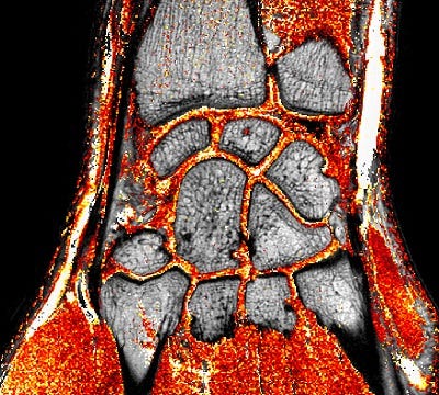
Figure 1: High resolution anatomical image with an overlay T1ρ-map in a healthy subject. The T1ρ-relaxation time is known to be sensitive to degeneration of cartilage, and is therefore used as a non-invasive biomarker for cartilage damage and repair.
The UMC Utrecht Center for Image Sciences participates actively in projects for which the direct detection of collagen can be used as a biomarker for fibrosis. Additionally, other techniques such as Chemical Exchange Saturation Transfer and Electrical Property Tomography can be used to monitor changes in tissue composition and physiology that play an important role in the delivery and follow up tissue regeneration and repair in cardiovascular and MSK related diseases. These MRI techniques have been developed for low as well as for ultra high field strengths. Besides the collaborative work with prof. Pieter Doevendans and dr. Steven Chamuleau in the area of cardiac tissue regeneration, we participate in a CVON consortium (1Valve) in collaboration with the TU Eindhoven (profs Frank Baaijens and Carlijn Bouten) focused on valve repair, as well as in a large European consortium funded by IMI (Innovative Medicine Innitiave; the Approach project) under UMCU leadership of profs Harrie Wijnans and Floris Lafeber in the area of osteoarthritis.

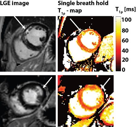
Figure 2: Native T1ρ-maps to detect myocardial fibrosis quantitatively without the use of a contrast agent. The corresponding Late Gadolinium Enhancement are shown for reference. Arrows indicate the infarcted area.
